Message on the topic of glass sponges biology. Sponges. Classes: lime, glass, ordinary. AT 4. The mantle cavity of mollusks is a cavity
Sponges are exceptionally peculiar Animals. Their appearance and body structure are so unusual that for a long time they did not know where to attribute these organisms to plants or animals. In the Middle Ages, for example, and even much later, sponges, along with other similar “doubtful” animals (bryozoans, some coelenterates, etc.), were placed among the so-called zoophytes, that is, creatures, as it were, intermediate between plants and animals. In the future, sponges were looked at either as plants or as animals. Only in the middle of the 18th century, when they became more familiar with the vital activity of sponges, was their animal nature finally proved. For a long time, the question of the place of sponges in the system of the animal kingdom remained unresolved. Initially, a number of researchers considered these organisms as colonies of protozoa, or unicellular animals. And this view seemed to be confirmed in the discovery by D. Clark in 1867 of choanoflagellates, flagellates with a plasma collar, which show a surprising resemblance to special cells - choanocytes, found in all sponges. However, soon after that, in 1874-1879, thanks to the studies of I. Mechnikov, F. IIIulze and O. Schmidt, who studied the structure and development of sponges, their belonging to multicellular animals was irrefutably proved.
Unlike a colony of protozoa, which consist of more or less monotonous and independent cells, in the body of multicellular animals, cells are always differentiated both in terms of structure and in terms of the function they perform. Cells here lose their independence and are only parts of a single complex organism. They form various tissues and organs that perform a specific function. Some of them serve for respiration, others perform the function of digestion, others provide excretion, etc. Therefore, multicellular animals are sometimes also called tissue animals. In sponges, the cells of the body are also differentiated and tend to form tissues, however, very primitive and weakly expressed. Even more convincing is the fact that sponges belong to multicellular animals that they have a complex individual development in their life cycle. Like all multicellular organisms, sponges develop from an egg. The fertilized egg divides many times, resulting in an embryo, the cells of which are grouped in such a way that two different layers are formed: the outer (ectoderm) and the inner (endoderm). These two layers of cells, called germ layers or sheets, with further development form strictly defined parts of the body of an adult animal.
After sponges were recognized as multicellular organisms, several more decades passed before they took their real place in the animal system. For quite a long time, sponges were classified as intestinal animals. And although the artificiality of their association with coelenterates was obvious, only since the end of the last century, the view of sponges as an independent type of the animal kingdom began to gradually win universal recognition. This was largely facilitated by the discovery by I. Delazh in 1892 of the so-called "perversion of the germ layers" during the development of sponges - a phenomenon that sharply distinguishes them not only from coelenterates, but also from other multicellular animals. Therefore, at present, many zoologists tend to subdivide all metazoans (Metazoa) into two divisions: Parazoa, to which only one type of sponge belongs to modern animals, and Eumetazoa, covering all other types. According to this idea, Parazoa includes such primitive multicellular animals whose body does not yet have real tissues and organs; in addition, in these animals the germ layers change places in the process of individual development, and in one way or another similar parts of the body of an adult organism, compared with Eumetazoa, arise in them from diametrically opposite rudiments.
Thus, sponges are the most primitive multicellular animals, as evidenced by the simplicity of their body structure and lifestyle. These are aquatic, predominantly marine, immobile animals, usually attached to the bottom or various underwater objects.
APPEARANCE OF SPONGE AND STRUCTURE OF THEIR BODY
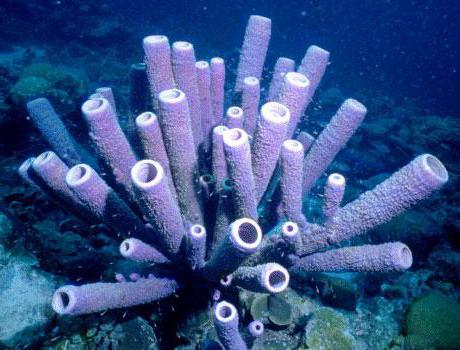
The body shape of sponges is extremely diverse. They often appear as crusty, cushion-like, carpet-like, or lumpy growths and outgrowths on stones, mollusc shells, or some other substrate. Often among them there are also more or less regular spherical, goblet-shaped, funnel-shaped, cylindrical, stalked, bushy and other forms.
The surface of the body is usually uneven, needle-like or even bristly to varying degrees. Only sometimes it is relatively smooth and even. Many sponges have a soft and elastic body, some are more rigid or even hard. The body of sponges is distinguished by the fact that it is easily torn, broken or crumbled. Having broken the sponge, one can see that it consists of an uneven, spongy mass, penetrated by cavities and channels running in different directions; elements of the skeleton - needles or fibers - are also quite well distinguishable.
The sizes of sponges vary widely: from dwarf forms, measured in millimeters, to very large sponges, reaching one meter in height or more.
 ,
,  ,
, 
Many sponges are brightly colored: most often in yellow, brown, orange, red, green, purple. In the absence of pigments, the sponges are white or gray in color.
The surface of the body of sponges is pierced by numerous small holes, pores, from which the Latin name of this group of animals comes from - Porifera, that is, porous animals.
With all the variety of appearance of sponges, the structure of their body can be reduced to the following three main types, which received special names: ascon, sicon and leukon.

Ascon. In the simplest case, the body of the sponge looks like a small thin-walled goblet or pouch, the base attached to the substrate, and the opening, which is called the mouth or osculum, facing upwards. The pores penetrating the walls of the body lead to a vast internal, atrial, or paragastric, cavity. The walls of the body consist of two layers of cells - the outer and the inner. Between them is a special structureless (gelatinous) substance - mesoglea, which contains various kinds of cells. The outer layer of the body consists of flat cells - pinacocytes, which form the covering epithelium, which separates the mesoglea from the water surrounding the sponge. Separate larger cells of the covering epithelium, the so-called porocytes, have an intracellular channel that opens outward with a pore opening and provides a connection between the internal parts of the sponge and the external environment. The inner layer of the body wall consists of characteristic collar cells, or choanocytes. They have an elongated shape, equipped with a tourniquet, the base of which is surrounded by a plasma collar in the form of an open funnel facing the atrial cavity. The mesoglea contains immobile stellate cells (collenocytes), which are connective tissue supporting elements, skeletal-forming cells (scleroblasts), which form the skeletal elements of sponges, various kinds of mobile amoebocytes, as well as archeocytes - undifferentiated cells that can turn into all other cells, and in addition number in sex. This is how sponges of the simplest asconoid type are arranged. Choanocytes here line the atrial cavity, which communicates with the external environment through pores and mouths.
Seacon. Further complication in the structure of the sponges is associated with the growth of the mesoglea and the invagination of parts of the atrial cavity into it, forming radial tubes. Choanocytes are now concentrated only in these invaginations, or flagellar tubes, and disappear from the rest of the atrial cavity. The walls of the body of the sponge become thicker, and then special passages are formed between the surface of the body and the flagellated tubes, which are called adductor canals. Thus, with a syconoid type of sponge structure, choanocytes line flagellar tubes that communicate with the external environment, on the one hand, through external pores or a system of adducting canals, and on the other, through the atrial cavity and orifice.
lacon. With even greater growth of the mesoglea and immersion of choanocytes into it, the most developed, leuconoid type of sponge structure is formed. Choanocytes are concentrated here in small flagellar chambers, which, unlike the sicon-type flagellar tubes, do not open directly into the atrial cavity, but are connected with it by a special system of discharge channels. Consequently, with the leukonoid type of sponge structure, choanocytes line flagellar chambers that communicate with the external environment, on the one hand, through external pores and adductor canals, and on the other, through a system of discharge canals, the atrial cavity and orifice. Most adult sponges have a leuconoid body type. In leucone, as well as in sicon, the covering epithelium (pinacocytes) lines not only the outer surface of the sponge, but also the atrial cavity and the canal system.
It should be borne in mind, however, that sponges in the process of growth often experience various kinds of complications in the structure of the body. The covering epithelium, with the participation of elements of the mesoglea, often thickens, turning into a dermal membrane, and sometimes into a cortical layer of different thicknesses. Extensive cavities form in places under the dermal membrane, from where the adducting channels originate. The same cavities can also form under the gastric membrane lining the atrial cavity. The exceptional development of the body of the sponge, its mesoglea, leads to the fact that the atrial cavity turns into a narrow canal and is often indistinguishable from the outlet canals at all. The system of flagellar chambers, canals, and additional cavities becomes especially complex and intricate when the sponges form colonies. At the same time, some simplifications can be observed associated with the almost complete disappearance of mesoglea in the body of sponges and the appearance of syncytia - multinuclear formations resulting from cell fusion. The covering epithelium may also be absent or replaced by syncytium.
In addition to the cells noted above, in the body of sponges, especially near numerous holes, cavities and channels, there are also special spindle-shaped myocyte cells capable of contraction. In some sponges, stellate cells were found in the mesoglea, interconnected by processes and sending processes to choanocytes and cells of the covering epithelium. These stellate cells are considered by some investigators as nerve elements capable of transmitting stimuli. It is quite possible that such cells play some kind of connecting role in the body of sponges, but there is no need to talk about the transmission of impulses that distinguish nerve cells. Sponges react very weakly even to the strongest external stimuli, and the transmission of stimuli from one part of the body to another is almost imperceptible. This indicates the absence of a nervous system in sponges.
Sponges are so primitive multicellular animals that the formation of tissues and organs in them is in its most infancy. For the most part, sponge cells have considerable independence and perform certain functions independently of one another, without connecting with each other into any tissue-like formations. Only the layer of choanocytes and the covering epithelium form something like tissues, but even here the connection between cells is extremely insignificant and unstable. Choanocytes can lose flagella and go into the mesoglea, turning into amoeboid cells; in turn, amoebocytes, rearranging, give rise to choanocytes. Covering epithelial cells also, plunging into the mesoglea, can turn into amoeboid cells.
BASIC LIFE DEPARTMENTS OF SPONGES.
As already noted, sponges are motionless animals and are not capable of any changes in the shape of the body. Only with a fairly strong irritation in some sponges, a very slow narrowing of the openings (mouths and pores) and channel lumen is observed, which is achieved by contraction of myocytes or protoplasm of other cells. Observations of shallow-water sponges living in the tidal zone have shown, for example, that their mouths close in 3 minutes and fully open in 7-10 minutes.
Most of the cells in the body of sponges are capable of releasing and retracting prolegs, or pseudopodia, or even using them to move through the thickness of the mesoglea. Amebocytes are especially mobile, sometimes moving at a speed of up to 20 microns per minute. But the most active cells of sponges are choanocytes. Their flagella are in constant motion. Due to the coordinated helical vibrations of the flagella of many choanocytes, a current of water is created inside the sponge. Water enters through the pores and the system of adducting channels into the flagellar chambers, from where it is directed through the outlet system of channels into the atrial cavity and is brought out through the mouth. Naturally, in sponges of a syconoid, and especially asconoid type of structure, the path of water is significantly reduced. It is very good to observe this flow of water in an aquarium, if a small amount of finely ground carcass is released near the sponge living there. You can see how the paint particles are carried into the sponge through the pores, and after a while they come out. An even more vivid picture is observed if a certain amount of carmine is injected into the body of the sponge with a syringe. Very soon, fountains of red liquid begin to beat from the nearest mouth openings.
The presence in the channel system of sponges of constant movement of water plays an important role in their life.
Breath. Like most animals living in the aquatic environment, sponges use oxygen dissolved in water for breathing. The current of water penetrating into all the cavities and channels of the sponge supplies the nearby cells and mesoglea with oxygen and carries away the carbon dioxide they release. Thus, gas exchange with the external environment is carried out in sponges directly by each cell or through the mesoglea.
Food. Sponges feed mainly on the remains of dead animals and plants suspended in water, as well as small unicellular organisms. The size of food particles usually does not exceed 10 microns. Food particles are brought with a current of water to the flagellar chambers, where they are captured by choanocytes and then enter the mesoglea. Here the food gets to the amoebocytes, which carry it to all parts of the body of the sponge. Inside these cells, in the digestive vacuoles that form around the trapped particles, food is digested. This process of digestion in sponges can be observed directly under a microscope. It can be seen how the amebocyte forms an outgrowth of the body - a pseudopod, directed towards the food particle entering the mesoglea. Gradually, the pseudopod covers this particle and draws it into the cell. Already in the elongated pseudopod, a digestive vacuole appears - a vesicle filled with liquid contents, which first have an acidic and then an alkaline reaction, in which food is digested. The captured particle dissolves, and grains of a fat-like substance appear on the surface of the vacuole. This is how the digestion and assimilation of food material by sponge cells occurs. Larger particles that get stuck in the adductor canals are captured by the cells lining them and also enter the mesoglea. If such a particle is too large and does not fit inside the amoeboid cell, it is surrounded by several amoebocytes, and digestion of food occurs inside such a cell mass. In some sponges, food digestion also occurs in choanocytes.
Selection. Undigested food residues are thrown into the mesoglea and gradually accumulate near the outlet canals, and then enter the lumens of the canals and are brought out. Sometimes the amoebocytes themselves, approaching the discharge canals, secrete the granular contents of their vacuoles there.
Sponges do not have the selective ability to capture only food particles. They absorb everything suspended in the water. Therefore, a large number of small inorganic particles constantly enter the body of the sponge. Their further fate is quite eloquently evidenced by the experience of coloring the water of an aquarium with carmine. Very soon, red particles of carmine get inside the choanocytes, and then into the mesoglea, where they are picked up by amoebocytes. Gradually, the entire sponge turns red, and its cells overflow with particles of carmine. After a few days, the cells of the sponge, and primarily choanocytes, are freed from these inorganic particles and the sponge acquires a normal color.
Consequently, the basic vital functions of sponges are carried out in an extremely primitive way. In the absence of special organs, the processes of respiration, nutrition and excretion proceed intracellularly, due to the activity of individual cells. It can be said that the level of physiology of sponges in this respect is only slightly higher than the level of physiology of unicellular animals.
SKELETON AND CLASSIFICATION OF SPONGE
Sponges almost always have an internal skeleton that supports the entire body and the walls of numerous channels and cavities. The skeleton can be calcareous, silicon or horny. The mineral skeleton consists of many needles, or spicules, which have a variety of shapes and are located in various ways in the body of sponges. Among the needles, macrosclera, which make up the bulk of the skeleton, and smaller and differently arranged microsclera are usually distinguished. Macrosclerae are mainly represented by simple, or uniaxial, three-beam, four-beam and six-beam needles. In the formation of the skeleton, in addition to the needles, a special organic substance, spongin, often takes part, with the help of which individual needles stick together with each other. Sometimes adjacent needles are soldered by the ends, making up the lattice skeletal frame of sponges of different strength. In the absence of mineral formations, the skeleton can be formed by horny (spongin) fibers alone.
The classification of sponges is largely based on skeletal structure. The substance from which the needles are formed, their shape and the general plan of the skeleton structure are taken into account. Each type of sponge contains characteristic needles of one or more often several varieties, differing in shape and size.
Sponges are divided into three classes: calcareous(Calcispongia) glass, or six-beam(Hyalospongia), and ordinary sponges (Demospongia). The former include sponges with a calcareous skeleton, the latter contain six-beamed silicon needles, and the latter include all the others, i.e. sponges with four-beam and uniaxial silicon needles, as well as horn sponges and very few sponges completely devoid of a skeleton.
Type PORIFERA
Class Calcispongia, or Calcarea
Order Homocoela
Order Heterocoela
Class Hyalospongia, or Hexactinellida
Order Hexasterophora
Order Amphidiscophora
Demospongia class
Order Tetraxonida
Order Cornacuspongida
REPRODUCTION AND DEVELOPMENT OF SPONGES
The reproduction of calcareous, silicohorn, and partly four-beam sponges has been best studied. Regarding glass sponges, quite reliable information is available only about their asexual reproduction.
sexual reproduction. Among the sponges, both dioecious and hermaphroditic forms are found. There is no external difference between males and females in the case of dioeciousness. Sex cells are formed from archeocytes in the mesoglea of the sponge. There is also the growth and maturation of eggs and the formation of spermatozoa. Mature spermatozoa come out of the sponge and with the flow of water through the system of adducting canals enter the flagellar chambers of other sponges that have mature eggs. Here they are captured by choanocytes and transferred to the mesoglea by amoebocytes, which transport them to the eggs. Sometimes the choanocytes themselves, losing flagella, like amoebocytes, transfer spermatozoa to the eggs, usually located near the flagellar chambers.
The crushing of the egg and the formation of the larva in most sponges proceed inside the mother's body. Only in representatives of some genera of four-beam sponges (Cliona, Tethya) do eggs go outside, where they develop.
The sponge larva, as a rule, has an oval or round body shape up to 1 mm in size. Its surface is covered with flagella, thanks to the movement of which the larva energetically swims in the water column. The duration of the free swimming of the larva until the moment of its attachment to the substrate varies from several hours to three days. In most sponges, the floating larva consists of an internal (mesogleal) mass of loosely arranged large granular cells, covered on the outside with a layer of smaller cylindrical flagellated cells. Such a two-layer larva is called a parenchymula and occurs as a result of uneven and improper crushing of the egg. Already at the first stages of crushing, cells of various sizes are formed: macro- and micromeres. Rapidly dividing micromeres gradually overgrow a compact mass of larger macromeres, and thus a two-layer parenchymula larva is obtained. In calcareous sponges (Homocoela) and in some of the most primitive four-beam sponges(Plakina, Oscarella) the larva initially looks like a vesicle, the shell of which consists of a single layer of homogeneous prismatic cells equipped with flagella. This larva is called a coeloblastula. Upon leaving the mother's body, it undergoes some metamorphosis, which consists in the fact that part of the cells, losing flagella, sinks into the larva, gradually filling the cavity existing there. As a result, the coeloblastula larva turns into the parenchymula already known to us. At the other part lime sponges(Heterocoela) larva also has the appearance of a single-layer vesicle, but differs in that its upper half (front part) is formed by small cylindrical cells equipped with flagella, and the lower (back) consists of large rounded granular cells. A similar single-layer larva, consisting of two types of cells, is called an amphiblastula. It retains this appearance until it is attached to the substrate.
Thus, sponges have two main larval forms: parenchymula and amphiblastula. After swimming for some time, the larva settles on a suitable substrate, attaching itself with its front end, and gradually a young sponge is formed from it. At the same time, in the parenchymula, a very interesting process, which is characteristic only of sponges, is observed of the movement of germ layers, which change their places. The flagellar cells of the outer ectodermal layer of the larva migrate into the inner cell mass, turning into choanocytes that line the resulting flagellar chambers. Endoderm cells lying under the outer layer of the larva, on the contrary, appear on the surface and give rise to the integumentary layer and mesoglea of the sponges. This is the so-called "germ perversion in sponges.
Nothing similar is observed in other multicellular animals: from the ectoderm and endoderm of their larvae, respectively, the ectodermal and endodermal formations of the adult organism are formed.
The development of sponges, which have a floating amphiblastula larva, proceeds somewhat differently. Before attaching such a larva to the substrate, its anterior hemisphere with small ectodermal flagellar cells protrudes inward, and the embryo becomes two-layered. The larger endodermal cells of the amphiblastula form the outer layer of the sponge, and the choanocytes of the flagellar chambers are formed due to the flagellar cells. As can be seen, in this case there is also a perversion of the germ layers. In other multicellular animals that have a vesicular larval stage (blastula) in development, consisting of cells of various sizes, larger cells give rise to the endoderm of an adult animal, and small cells (forward hemisphere) give rise to ectoderm. In sponges, we observe just the opposite relationship. As a result of the larval metamorphosis of sponges, accompanied by the formation of an atrial cavity, an orifice, and skeletal elements, post-larval stages are obtained - olintusiliragon. Olynthus is a small bag-shaped sponge of an asconoid type of structure. With its further growth, single or colonial calcareous Homocoela are formed, and with a corresponding complication of the structure, others lime sponges(Heterocoela). Ragon is characteristic of ordinary sponges. It has the appearance of a sponge of a syconoid structure, strongly flattened in the vertical direction, with an extensive atrial cavity and an orifice at the apex. After some time, the ragon transforms into a young sponge of the leuconoid type. Interestingly, some representatives ordinary sponges(Halisarca), like calcareous sponges, in their development they first go through a stage that has the most primitive, asconoid type of structure. It is impossible not to see in this a manifestation of the biogenetic law, according to which animals in their individual development successively pass through certain stages corresponding to the main features of the structure of their ancestors.
asexual reproduction. In sponges, various forms of asexual reproduction are very widespread. These include external budding, the formation of sorites, gemmules, etc.

When budding, the separated daughter individual may contain all the tissues of the mother's organism and be simply its isolated section. Similar budding is observed in calcareous, as well as in some glass and primitive four-ray sponges. In other cases, the kidney arises from a collection of archaeocytes. The most famous example of such kidney formation is sea orange(Tethya anrantium). A group of archeocytes accumulates on the surface of the sponge; a small sponge is gradually formed from them, which after a while is laced from the mother's body, falls off and proceeds to an independent lifestyle. Sometimes the kidneys are formed at the ends of the needles protruding from the body of the sponges, or a beaded series of small kidneys connected in series with each other, which have a weak connection with the mother's organism, is formed. As an extreme case of such budding, a special method of asexual reproduction observed in some geodia(Geodia barretti). Archeocytes here extend beyond the maternal sponge; at the same time, some long needles are pushed out of it, settling to the bottom. On these needles, as on a substrate, a reproducing mass of archeocytes accumulates, and quite independently of the maternal organism, a small sponge of geodia is gradually formed. The formation of external buds from accumulations of archaeocytes is widespread in many four-beam sponges(Tethya, Polymastia, Tetilla, etc.) "and is also sometimes found in silicon horn and other sponges.
Much less often, asexual reproduction is observed in sponges, which is expressed in the separation from the mother's organism of various sizes of areas, which then develop into an adult organism. Very closely adjacent to this method of reproduction is the formation in sponges under unfavorable conditions for the existence of the so-called reduction bodies. This process is invariably accompanied by the disintegration of a significant part of the body of the sponge. The surviving part is isolated in the form of several cell clusters, or reduction bodies, consisting of a group of amoebocytes, dressed on the outside with cells of the covering epithelium. With the onset of favorable conditions, new sponges develop from these reduction bodies. The formation of reduction bodies occurs in marine sponges, especially those living in the tidal zone, as well as in freshwater sponges that do not have the ability to form gemmules.
It is also characteristic of sponges to reproduce with the help of sorites and gemmules. This method of reproduction is sometimes called internal budding. Sorites are rounded formations, much less than 1 mm in diameter. They arise inside the sponges from the accumulation of archeocytes. During the development of the sorite, the embryo usually forms from a single cell that feeds on the remaining cells of the sorite, which have merged into a syncytial mass. Sorites can produce free-swimming larvae that are essentially no different from those formed sexually. Such a larva subsequently undergoes metamorphosis and turns into a young sponge. Sorites are known in many common sponges, including the freshwater Baikal sponge. It is easy to see that asexual reproduction with the help of sorites is extremely close to the sexual parthenogenetic reproduction observed in some multicellular animals. In sponges, therefore, there is an extreme convergence of the phenomena of asexual and sexual reproduction. This is due to the fact that undifferentiated amoeboid cells, or archeocytes, not only give rise to germ cells, but also take part in various forms of asexual reproduction.

Gemmules, like sorites, are formed inside sponges from accumulations of archeocytes. They are very characteristic of freshwater sponges and often have a rather complex structure. In the formation of gemmules, a group of nutrient-rich archaeocytes is surrounded by a dense chitinous capsule, and then an air-bearing layer containing usually special gemmulus microsclerae, which are often located on the surface of the capsule in regular rows. Typically, the capsule is provided with a small hole for the exit of its contents to the outside. Gemmules are a dormant stage and can persist for a long time under adverse conditions that cause the death of the sponge itself. In a living or dead sponge, such gemmules, about 0.3 mm in diameter, are sometimes found in very large numbers. When favorable conditions occur, differentiation of cells begins in the gemmules, which come out in the form of a shapeless mass and then form a young sponge. Gemmules are also found in some marine sponges (Suberites, Cliona, Haliclona, Dysidea, etc.) ”but here they are simpler in structure than in badyags, and do not have special skeletal elements.
SPONGE-COLONIAL ORGANISMS
Relatively few sponges are solitary organisms. These are, for example, lime sponges various types of structure, with one mouth at the top, as well as glass and some ordinary sponges fairly regular and symmetrical body shape. In general, any sponge that has one mouth and a single channel system associated with it is considered as a single organism. Most of the sponges, however, are represented by various kinds of colonial formations. It is generally accepted that colonies arise as a result of asexual reproduction that has not been completed. It can be imagined that on the surface of a single sponge, small sponges are formed by budding, which do not separate from the mother organism. They continue to exist together, forming a colony that unites a different number of individuals, or individuals.

Such colonies do occur in some calcareous(Leucosolenia, Sycon, etc.) and glass sponges(Rhabdocalyptus, Sympagella, etc.), which form small groups of individuals connected by their bases. But usually in sponge colonies, individual individuals merge with each other to varying degrees. This fusion occurs very early and is often so complete that it is difficult, even impossible, to distinguish the individuals of the colony from each other. On the surface of the colony, in such cases, only the mouth opening is preserved from each individual. Therefore, it is conditionally customary to consider in such colonies a part of the body of a sponge with one mouth as a separate individual. The formation of this kind of colonies is affected by the great simplicity of the structure of the sponges, the low level of integrity and the weakly expressed individuality of their organism. Not only the individual individuals that make up the colony, but often the colonies themselves, which look like shapeless formations, are distinguished by an exceptionally weak individuality. These are cortical, lumpy, stalked, bushy and other sponges of irregular and indefinite body shape, characterized by great variability in appearance. They are especially indicative of silicon-horn and four-beam sponges. It is characteristic that such colonies can be formed not only from one individual of the sponge, but also by the merger of sponges of the same species growing nearby. Moreover, even their larvae are able to join together and give rise to a colony.
The situation is different when the sponge acquires a definite or regular body shape. The mouths, which serve to recognize individual individuals of the colony, here represent formations that, to one degree or another, are subordinate to the sponge as a formalized whole. There is a process of individualization of the colony with complete dissolution of individual individuals in it. This is observed in many four-rayed and some silicon-horned sponges, which have a more or less regular and symmetrical body shape. Such are, for example, cup-shaped, goblet-shaped or funnel-shaped forms, often equipped with a stem. Their mouth openings are located on the inner surface of the funnel, and the pores are outside. These sponges are already formations of a higher order than ordinary shapeless colonies. But the process of individualization of the colony can go further. The edges of the glass or funnel, stretching upward, grow together in such a way that a cavity (pseudo-atrial) is formed inside the sponge, opening at the top with a hole, which now functions as a single mouth. And outwardly, such a tubular or bag-shaped sponge resembles many single glass sponges. A similar process is observed in spherical or similar forms. The mouths here can be scattered over the entire surface, collected in various ways in small groups, or even merged into one hole. In the latter case (as, for example, in some representatives of the family Tetillidae, Geodiidae, etc.), there are no traces of the former coloniality. From the very beginning of development, such regular forms grow as a whole. Here we have an example of the emergence of secondarily single individuals of sponges. Such single sponges are also found among cushion-shaped and disc-shaped representatives of sponges - polymastium(family Polymastiidae), which have one estuarine papilla on the surface, and in a number of other four-beam sponges. Very close to them are highly individualized colonies of silicon horn sponges, which have a regular radially symmetrical body shape. Such are many sponges sea brushes, tubular, funnel-shaped and other forms. But the individuality of such sponges is very imperfect and unstable. Often already secondary single forms form additional mouths, thereby showing their original colonial essence. A sponge is a good example of this phenomenon. sea mushroom, at the top of which there is one mouth. Such a sponge is a secondary single organism.
Under certain conditions, however, specimens with two or more mouths appear. The same can be observed in sponges from the Tetillidae family.
Thus, ordinary sponges are mainly represented by either formless colonial formations or secondary single individuals and transitional forms between them in the form of highly individualized colonies. Lime and glass sponges contain a certain number of solitary forms, they can form various kinds of colonies, including well-isolated individuals.
SPONGE REGENERATION PHENOMENON
Regeneration refers to the restoration of lost parts of the body by the body. Many animals are capable of regeneration, and the simpler the organism is arranged, the more powerful this ability is manifested. It is known, for example, that a hydra can be cut into many pieces and a new hydra is restored over time from each piece. Sponges have even greater potential for regeneration. This is evidenced by the classical experiments of G. Wilson on the restoration of sponges from individual cells. If pieces of sponges are rubbed through a fine-mesh cloth, the result is a filtrate containing isolated cells. These cells remain viable for several days, showing a brisk amoeboid movement, that is, releasing pseudopods and moving with their help. Placed at the bottom of the vessel, they gather in groups, forming shapeless clusters, which after 6-7 days turn into small sponges. Interestingly, when mixing filtrates obtained from different sponges, only homogeneous cells come together, forming sponges of the corresponding type.
Undoubtedly, the experiments cited equally, if not more, also characterize the process of artificially induced asexual reproduction of sponges, which, as we already know, very often occurs through the accumulation of reproducing cell masses. And this is a feature of the regenerative processes in sponges. They are so closely intertwined with the phenomenon of asexual reproduction that it is sometimes difficult to draw clear boundaries between them, just as it is sometimes difficult to distinguish between normal growth and reproduction by budding. This is especially true of colonial sponges that do not have a definite body shape.
Therefore, often when the sponge is damaged, the process that began as a recovery process ends with asexual reproduction. So, we observed how on the surface of the sponge sea loaf(Halichondria panicea) at the site of a deep wound, during the restoration of damaged parts, numerous mouth openings and papilles were formed. It is also known that, under certain conditions, it is possible to artificially induce bud formation in calcareous and freshwater sponges as a result of mechanical damage or burns.
In its purest form, the regeneration process can be observed on single sponges or in case of damage to any formed formations (mouth tubes, or papillae, dermal membrane) on the body of colonial sponges. In general, sponges quite easily regenerate damaged areas on the surface of the body. The wound quickly cicatrizes, tightens with a membrane, and the previous structure is restored, so that very soon the place of damage becomes invisible. A shallow incision through a sea loaf sponge, for example, is eliminated after 5-7 days, and a hole of about 1 sq. mm, done near the mouth lime sponge(Leucosolenia), overgrown in 7-10 days. However, with more significant damage, the recovery process often proceeds very slowly. So, if the upper part bearing the mouth is cut off from a single calcareous sponge (Sycon), then the dermal membrane regenerates at the remaining base of the sponge in a day and a new mouth is formed; but only after 15 days flagellar tubes are formed here. With a more significant and deeper damage to the body of the sponge sea loaf, healing also proceeds rather slowly and, moreover, often recovery is not complete. Obviously, the great regenerative capacity of sponges cannot be sufficiently manifested here, in view of the fact that the integrity, or degree of integration, of the sponges themselves as multicellular animals is still extremely insignificant.
When the sponge is cut into two parts and then closely connected, these parts grow together very quickly. Pieces taken from different specimens of the same species can also grow together. It is characteristic that in some cases, when the cut has passed through the mouth papilla, during fusion, two small papillae are formed instead of one, i.e., regeneration ends with the formation of a new individual of the colony. A living sponge can be cut into many pieces, and each piece remains alive. On its damaged surface, the dermal membrane is restored, a mouth is formed, and each cut piece continues its existence and growth in the future, like a whole sponge.
The method of artificial breeding of commercial toilet sponges is based on the ability of sponges to regenerate. Such a sponge is cut into pieces and attached with a wire to some solid substrate. Most often, special cement discs are used for this, which are placed at the bottom. Sometimes pieces of sponge are strung on a cord stretched horizontally at the very bottom between two stakes. After a few years, a specimen grows from a piece of sponge, reaching a commercial size. True, they note that with a similar method of reproduction, the sponge grows much more slowly than when it develops from a larva.
SPONGE LIFETIME
The lifespan, or age that sponges reach, varies between species from a few weeks and months to many years. Lime sponges usually live up to one year on average. Some of them (Sycon coronatum, Grantia compressa) die off immediately upon reaching puberty, as soon as the formed larvae of the new generation leave their body. Most small four-beam and silicon-horned sponges live within 1-2 years. Larger glass and ordinary sponges are long-lived organisms. Particularly durable are those that reach a value of 0.5 m or more. Yes, instances horse sponge(Hippospongia communis) about 1 m in diameter, according to experts, reach an age of at least 50 years.
In general, sponges grow rather slowly. The highest growth rate in forms with a short lifespan. Some lime sponges(Sycon ciliatum) in 14 days grew up to 3.5 cm in height, i.e., they reached almost their maximum size. detached kidney sea orange acquires the size of the mother's organism (2-3 cm in diameter) within one month. On the contrary, a long-lived horse sponge grows up to 30 cm in diameter in 4-7 years. It must be assumed that other large sea sponges have approximately the same growth rate. Of course, in each individual case, the growth rate and life span of sponges largely depend on various environmental factors, including the abundance of food, temperature conditions, etc.
Freshwater sponges are relatively short-lived and usually only live for a few months. But in some cases they are able to create perennial formations of a special kind. Such formations, reaching a significant size and weighing more than 1 kg, consist of an internal mass of dead parts of the sponge, covered on the outside with a vital layer. It happens in the following way. The sexually produced sponge larva attaches itself to a suitable substrate and grows into a small colony. Having formed gemmules, such a sponge dies. After some time, with the onset of favorable conditions, young sponges emerge from the gemmules. They rise to the surface of the dead sponge and, merging with each other, form a young colony. Such a restored colony, having reached a certain age, proceeds to sexual reproduction. Later, new gemmules form inside it, and the sponge itself dies. The following year, the cycle repeats itself, and in this way voluminous colonies of freshwater sponges can gradually be created.
Biological Encyclopedic Dictionary Geological Encyclopedia Wikipedia
Animals Clockwise from upper left: European squid (mollusks), sea nettle (cnidaria), leaf beetle (arthropod), nereid (annelid worms) and tiger (chordates) ... Wikipedia
Sponges are ancient primitive multicellular animals. They live in marine, less often fresh water bodies. They lead a fixed lifestyle. They are filter feeders. Most species form colonies. They do not have tissues or organs. Almost all sponges have an internal skeleton. The skeleton is formed in the mesoglea and can be mineral (calcareous or silicic), horny (sponginous) or mixed (silicic-sponginous).
There are three types of sponge structure: ascon (asconoid), sicon (syconoid), leukon (leuconoid)
The most simply organized sponges of the asconoid type are in the form of a bag, which is attached to the substrate with its base, and the mouth (osculum) is turned upwards.
The outer layer of the sac wall is formed by integumentary cells (pinacocytes), the inner layer by collar flagellar cells (choanocytes). Choanocytes perform the function of water filtration and phagocytosis.
Between the outer and inner layers there is a structureless mass - mesoglea, in which there are numerous cells, including those forming spicules (needles of the internal skeleton). The entire body of the sponge is permeated with thin canals leading to the central atrial cavity. Continuous work of choanocyte flagella creates a water flow: pores → pore channels → atrial cavity → osculum. The sponge feeds on those food particles that the water brings.
Sponge structure
The body of the sponges is porous, has a goblet shape. In the upper part of the body there is a hole - the mouth through which the body cavity of the sponge (paragastric cavity) communicates with the environment. The body wall consists of outer (ectoderm) and inner (endoderm) layers, between which there is a gelatinous substance - mesoglea.

Mouth - an opening in the body of sponges through which the body cavity communicates with the environment.
Ectoderm is the outer layer of cells.
In the ectoderm are cells of the integumentary epithelium (squamous cells). Integumentary epithelium is a tissue consisting of cells tightly pressed to each other, separating the body from the environment, and performing barrier and protective functions.
Endoderm is the inner layer of cells. The endoderm consists of cells that have a flagellum - choanocytes. The movement of the flagellum ensures the injection of water into the body of the sponge.
Mesoglea is a gelatinous substance that is located between the outer and inner layers of the body. In the median gelatinous substance there are supporting cells that form the skeleton, amoebocytes that digest food and provide regeneration, as well as germ cells.
Regeneration is the restoration of lost parts of the body. The skeleton of sponges consists of needles - spicules, a substance - spongin, which fastens the needles together, takes part in its formation.
Spicules - needles that form the skeleton of sponges, mostly calcareous or silicic.
Spongin is the substance that holds the spicules together.
Nutrition, respiration and excretion
Nutrition, respiration and excretion of sponges is carried out due to the flow of water through their body. Water enters the pores and exits through the mouth. Nutrients from the water are taken up by choanocytes and transferred to amoebocytes, which digest them. Thus, according to the method of nutrition, sponges are filter feeders. Thanks to this, sponges can clean the water in the pond.
reproduction
Sponges are able to reproduce both sexually and asexually.
Asexual reproduction is by budding or fragmentation. During fragmentation, the body of the sponge breaks up into parts, each of which gives rise to a new organism. During sexual reproduction, the sperm from one sponge enters the body of another with a stream of water. As a result, a larval stage is formed, which enters the environment, attaches to the substrate and turns into an adult sponge. The larval stage is necessary for the settlement of sponges.
Sponges have well-developed passive defenses, such as the presence of mineral needles, the production of toxic chemicals.
SPONGE TYPE (PORIFERA or SPONGIA)
Sponges are exceptionally peculiar Animals. Their appearance and body structure are so unusual that for a long time they did not know where to attribute these organisms to plants or animals. In the Middle Ages, for example, and even much later, sponges, along with other similar “doubtful” animals (bryozoans, some coelenterates, etc.), were placed among the so-called zoophytes, that is, creatures, as it were, intermediate between plants and animals. In the future, sponges were looked at, sometimes as plants, sometimes as animals.
Only in the middle of the 18th century, when they became more familiar with the vital activity of sponges, was their animal nature finally proved. For a long time, the question of the place of sponges in the system of the animal kingdom remained unresolved. Initially, a number of researchers considered these organisms as colonies of protozoa, or unicellular animals.
And such a view seemed to be confirmed in the discovery by D. Clark in 1867 of choanoflagellates, flagellates with a plasma collar, which show a surprising resemblance to special cells - choanocytes, found in all sponges. However, soon after that, in 1874-1879, thanks to the studies of I. Mechnikov, F. IIIulze and O. Schmidt, who studied the structure and development of sponges, their belonging to multicellular animals was irrefutably proved.
Unlike a colony of protozoa, which consist of more or less monotonous and independent cells, in the body of multicellular animals, cells are always differentiated both in terms of structure and in terms of the function they perform. Cells here lose their independence and are only parts of a single complex organism. They form various tissues and organs that perform a specific function.
Some of them serve for respiration, others perform the function of digestion, others provide excretion, etc. Therefore, multicellular animals are sometimes also called tissue animals. In sponges, the cells of the body are also differentiated and tend to form tissues, however, very primitive and weakly expressed.
Even more convincing is the fact that sponges belong to multicellular animals that they have a complex individual development in their life cycle. Like all multicellular organisms, sponges develop from an egg. The fertilized egg divides many times, resulting in an embryo, the cells of which are grouped in such a way that two different layers are formed: the outer (ectoderm) and the inner (endoderm). These two layers of cells, called germ layers or sheets, with further development form strictly defined parts of the body of an adult animal.
After sponges were recognized as multicellular organisms, several more decades passed before they took their real place in the animal system. For quite a long time, sponges were classified as intestinal animals. And although the artificiality of their association with coelenterates was obvious, only since the end of the last century, the view of sponges as an independent type of the animal kingdom began to gradually win universal recognition.
This was largely facilitated by the discovery by I. Delage in 1892 of the so-called "perversion of the germ layers" during the development of sponges - a phenomenon that sharply distinguishes them not only from intestinal, but also from other multicellular animals. Therefore, at present, many zoologists tend to subdivide all metazoans (Metazoa) into two divisions: Parazoa, to which only one type of sponge belongs to modern animals, and Eumetazoa, covering all other types.
According to this idea, Parazoa includes such primitive multicellular animals whose body does not yet have real tissues and organs; in addition, in these animals the germ layers change places in the process of individual development, and in one way or another similar parts of the body of an adult organism, compared with Eumetazoa, arise in them from diametrically opposite rudiments.
Thus, sponges are the most primitive multicellular animals, as evidenced by the simplicity of their body structure and lifestyle. These are aquatic, predominantly marine, immobile animals, usually attached to the bottom or various underwater objects.
APPEARANCE OF SPONGE AND STRUCTURE OF THEIR BODY
The body shape of sponges is extremely diverse. They often appear as crusty, cushion-like, carpet-like, or lumpy growths and outgrowths on stones, mollusc shells, or some other substrate. Often among them there are also more or less regular spherical, goblet-shaped, funnel-shaped, cylindrical, stalked, bushy and other forms.
The surface of the body is usually uneven, needle-like or even bristly to varying degrees. Only sometimes it is relatively smooth and even. Many sponges have a soft and elastic body, some are more rigid or even hard. The body of sponges is distinguished by the fact that it is easily torn, broken or crumbled. Having broken the sponge, one can see that it consists of an uneven, spongy mass, penetrated by cavities and channels running in different directions; elements of the skeleton - needles or fibers - are also quite well distinguishable.
The sizes of sponges vary widely: from dwarf forms, measured in millimeters, to very large sponges, reaching one meter in height or more.
Many sponges are brightly colored: most often in yellow, brown, orange, red, green, purple. In the absence of pigments, the sponges are white or gray in color.
The surface of the body of sponges is permeated with numerous small holes, pores, from which comes the Latin name of this group of animals - Porifera, that is, porous animals.
With all the variety of appearance of sponges, the structure of their body can be reduced to the following three main types, which received special names: askon, sikon and leukon.
Ascon. In the simplest case, the body of the sponge looks like a small thin-walled goblet or pouch, the base attached to the substrate, and the opening, which is called the mouth or osculum, facing upwards. The pores penetrating the walls of the body lead to a vast internal, atrial, or paragastric, cavity. The walls of the body consist of two layers of cells - the outer and the inner. Between them is a special structureless (gelatinous) substance; mesoglea, which contains various types of cells.
The outer layer of the body consists of flat cells - pinacocytes, which form the covering epithelium, which separates the mesoglea from the water surrounding the sponge. Separate larger cells of the covering epithelium, the so-called porocytes, have an intracellular channel that opens outward with a pore opening and provides a connection between the internal parts of the sponge and the external environment. The inner layer of the body wall consists of characteristic collar cells, or choanocytes. They have an elongated shape, equipped with a tourniquet, the base of which is surrounded by a plasma collar in the form of an open funnel facing the atrial cavity.
The mesoglea contains immobile stellate cells (collencites), which are connective tissue supporting elements, skeletal-forming cells (scleroblasts), which form the skeletal elements of sponges, various kinds of mobile amoebocytes, as well as archeocytes - undifferentiated cells that can turn into all other cells, including number in sex. This is how sponges of the simplest asconoid type are arranged. Choanocytes here line the atrial cavity, which communicates with the external environment through pores and mouths.
Seacon. Further complication in the structure of the sponges is associated with the growth of the mesoglea and the invagination of parts of the atrial cavity into it, forming radial tubes. Choanocytes are now concentrated only in these invaginations, or flagellar tubes, and disappear from the rest of the atrial cavity. The walls of the body of the sponge become thicker, and then special passages are formed between the surface of the body and the flagellated tubes, which are called adductor canals.
Thus, with the syconoid type of sponge structure, choanocytes line flagellar tubes that communicate with the external environment, on the one hand, through external pores or a system of adducting canals, and on the other, through the atrial cavity and mouth.
lacon. With even greater growth of the mesoglea and immersion of choanocytes into it, the most developed, leuconoid type of sponge structure is formed. Choanocytes are concentrated here in small flagellar chambers, which, unlike the sicon-type flagellar tubes, do not open directly into the atrial cavity, but are connected with it by a special system of discharge channels.
Consequently, with the leukonoid type of sponge structure, choanocytes line flagellar chambers that communicate with the external environment, on the one hand, through external pores and adductor canals, and on the other, through a system of discharge canals, the atrial cavity and orifice. Most adult sponges have a leuconoid body type. In leucone, as well as in sicon, the covering epithelium (pinacocytes) lines not only the outer surface of the sponge, but also the atrial cavity and the canal system.
It should be borne in mind, however, that sponges in the process of growth often experience various kinds of complications in the structure of the body. The covering epithelium, with the participation of elements of the mesoglea, often thickens, turning into a dermal membrane, and sometimes into a cortical layer of different thicknesses. Extensive cavities form in places under the dermal membrane, from where the adducting channels originate.
The same cavities can also form under the gastric membrane lining the atrial cavity. The exceptional development of the body of the sponge, its mesoglea, leads to the fact that the atrial cavity turns into a narrow canal and is often indistinguishable from the outlet canals at all. The system of flagellar chambers, canals, and additional cavities becomes especially complex and intricate when the sponges form colonies.
At the same time, some simplifications can be observed associated with the almost complete disappearance of mesoglea in the body of sponges and the appearance of syncytia - multinuclear formations resulting from cell fusion. The covering epithelium may also be absent or replaced by syncytium.
The body of sponges consists of many cells that perform various functions. However, unlike other multicellular animals, sponges do not have tissue differentiation. 1 - paragastric cavity, 2 - mouth, 3 - choanaocytes (collar cells of the endoderm), 4 - ectoderm, 5 - needles of the mineral skeleton, 6 - canal.
In addition to the cells noted above, in the body of sponges, especially near numerous holes, cavities and channels, there are also special spindle-shaped cells - myocytes, capable of contraction. In some sponges, stellate cells were found in the mesoglea, interconnected by processes and sending processes to choanocytes and cells of the covering epithelium.
These stellate cells are considered by some investigators as nerve elements capable of transmitting stimuli. It is quite possible that such cells play some kind of connecting role in the body of sponges, but there is no need to talk about the transmission of impulses that distinguish nerve cells. Sponges react very weakly even to the strongest external stimuli, and the transmission of stimuli from one part of the body to another is almost imperceptible. This indicates the absence of a nervous system in sponges.
Sponges are such primitive multicellular animals that the formation of tissues and organs in them is in its most infancy.
For the most part, sponge cells have considerable independence and perform certain functions independently of one another, without connecting with each other into any tissue-like formations.
Only the layer of choanocytes and the covering epithelium form something like tissues, but even here the connection between cells is extremely insignificant and unstable. Choanocytes can lose flagella and go into the mesoglea, turning into amoeboid cells; in turn, amoebocytes, rearranging, give rise to choanocytes. Covering epithelial cells also, plunging into the mesoglea, can turn into amoeboid cells.
The underwater world is so diverse and unique that sometimes it is even difficult to distinguish plants from animals. Such bizarre forms are the creatures living there. Large sea giants and very microscopic planktonic crustaceans, colorful and bright, predators and herbivores - an insane variety of species of living organisms. One of these amazing creatures are sponges, which will be discussed later.
general information
You can characterize the position of these animals in the following way:
- empire - Cellular;
- kingdom - Animals;
- subkingdom - Multicellular;
- type - Sponges.
To date, it is known that there are about 8 thousand species. 300 of them inhabit the expanses of the seas of our country.
Classification
The Sponge type combines all known representatives into four large classes.
- Calcarea, or Calcareous. Formed in the form of deposited calcium salts.
- Ordinary, or Kremnerogovye. The main representative is a badyaga.
- Glass (Six-beam). The class size is small.
- Coral - a very poor species class.
All of these sponges have their own characteristics not only external, but also internal structure, lifestyle and economic importance in human life.

External structure
Perhaps the most unusual in the whole characterization of the animals in question will be precisely the external appearance. The features of the external structure of the sponge are determined by the variety of body shapes that are characteristic of them. So, representatives of different classes can be in the form:
- glasses;
- bowls;
- tree structure.
The symmetry of the body in single forms is bipolar axial, in colonial forms it is mixed. Each individual has a special flat sole, with which it is attached to the bottom or other substrate. Sponges lead most often an immobile way of life.
On the upper side of the body is a special opening, which is called the "osculum". It serves to remove excess water from the internal cavity. Outside, the body is covered with a layer of cells - pinacoderm. They resemble in their structure the epithelial tissue of higher animals.
However, they also have distinctive features - the presence of wide pores. The structure of the sponge provides for the absorption of food particles not through the upper hole, but through numerous perforations penetrating the entire body, capable of contracting and expanding.
Under the outer layer are two more, which we will consider in more detail later. The color scheme of both single and colonial forms is quite diverse. There are the following types of coloring:
- gray;
- green;
- purple;
- yellow;
- white;
- red;
- brown;
- mixed.
The Sponge type brings the underwater world to life, making it even brighter, more colorful and attractive. Moreover, if we consider a single individual on the land surface, then it will have a very unattractive appearance: a brownish slippery lump resembling a raw liver, emitting a not very pleasant aroma.

The internal structure of representatives
Sponge body types are similar, whether solitary or attached to a colony. Immediately under the dermal outer layer of porous cells is a special intercellular substance that forms a fairly voluminous membrane. In it, the cells are located loosely, and their shape is different. The tissue is somewhat reminiscent of fatty tissue in higher terrestrial representatives. This structure is called mesohyl.
Under this layer is an internal cavity lined with a special row of cells. This is the gastric layer. All food goes here, and digestion takes place here. All waste products, together with excess water, are sent to the upper opening of the body and are brought out through it.
Also, the structure of the sponge necessarily includes a kind of skeleton. It is formed from lime, phosphorus, organic salts, which are produced in special mesochil cells. It not only gives the sponges a certain shape of the body, but is also important for the preservation of the internal cavity from mechanical damage.
The characteristic of the Sponge type will be incomplete if the main feature of these animals is not indicated - their body does not have tissues, but only includes layers of various shapes and forms. This is the main difference between the considered animals from all others.

The aquifer system of individuals is also interesting. It may be different for different classes. There are three main types of it:
- Ascon - all communication with the external environment is carried out through a system of tubes through which water moves into special cell-chambers. The most simplified aquifer system found in a few representatives.
- Seacon. A more advanced system, which includes a network of branched tubules and tubules that flow into special cell-cameras with flagella.
- Leikon - a whole network of osculums, this type of aquifer system is typical only for colonial forms. The most complicated option against the background of all the previous ones.
Sponges reproduce both sexually and asexually. Sex cells are formed in the mesohyl layer. Then the products go out through the pores of the body and with the flow of water enter the bodies of other sponges, where fertilization occurs. As a result, a zygote is formed, giving rise to a larva. The fry can be called differently: amphiblastula, parenchymula, celloblastula.
If we talk about that, it is based on the process of budding, that is, detachment with subsequent regeneration of the missing structures. For the most part, the Sponge type includes hermaphroditic animals.
Lifestyle Features
If we consider the whole variety of multicellular animals of the world, then sponges should be attributed to the most primitive stage in terms of organization. However, these are also the most ancient animals that appeared many thousands of years ago. During the evolution of their organization, little has changed, they retain their characteristics over time. The life form of representatives has two manifestations:
- single;
- colonial.
Most often, mass accumulations of sponges are found among coral reefs. There are both freshwater species (their minority) and oceans (the overwhelming majority of species).

The Sponge type includes animals that feed on small organisms or their remains. In the structure of their body there are special collar cells with flagella. They just capture the floating particles of food, directing them into the internal paragastric cavity of the body. Digestion takes place inside the cells.
According to the method of obtaining food, sponges can be called passive hunters. They sit lazily in an attached place, waiting for passing nutrient particles. And only when they are already very close, they capture them through the pores and direct them, together with the flow of water, into the body.
Some species are able to move, despite the fact that they still have soles for attaching to the substrate. However, their speed is so low that for the whole day the individual is unlikely to move further than a meter.
Variety of sponges
For such primitive representatives, it is quite impressive - after all, there are about 8 thousand species of them! And according to some modern data, this figure is already approaching 9 thousand. External diversity is explained by the difference in body shape, skeleton types and body color of individuals (or colonies).
Grade Glass Sponges
Glass sponges are very interesting in their external variety. They are not as numerous as others, but have an unusual skeleton. These are one of the largest individuals that the Sponge type includes. The general characteristics of the representatives of this class can be expressed in several points.
- The Latin name of the class is Hexactinellida.
- The skeleton is formed from silicon compounds, so it is very durable.
- Needle type body support, which is dominated by six-pointed structures.
- Larvae of parenchymula or coeloblastula species.
- Aquifer system of the leukon type.
- More often colonial than solitary forms.
- Sometimes up to 50 cm in height.
The most common representatives are such as:
- hyalonema siboldi;
- euplectella.

Class Ordinary, or Kremnerogovye, sponges
The Sponge type, a photo of whose representatives can be seen in this article, also includes the most numerous class in terms of the number of individuals - Kremnerogye, or Ordinary. They got their name for the features in the structure of the skeleton - it consists of silica and spongin. The hardness is quite gentle and easily destroyed. The shape of the spines of the skeleton is very diverse:
- asterisks;
- anchors;
- clubs;
- sharp needles and so on.
The most common freshwater representative is badyaga - a sponge used as an indicator of the purity of the reservoir. Outwardly unattractive, the color is brown-brown, sometimes dirty yellow. Used by man for various needs.
What other representatives are found among ordinary sponges?
- Mixils.
- Sea caravan.
- Baikal sponge.
- Marine brushes.
- Giant chondrocladia and others.
Class Lime sponges
It includes representatives with a strong and beautiful calcareous skeleton. They live only in the seas and oceans. The coloration is pale or completely absent. The spines of the skeleton may have about three rays. The main representatives: ascons, sicons, leukandry.
Class Coral sponges
The fewest representatives that outwardly resemble coral branches. This happens due to the formation of a powerful calcareous skeleton of different colors and patterned structure.
Representatives: Nicholson's geratoporella, merlia. In total, only six species of such animals have been described. For a long time they were not distinguished from the coral reef system, so they were discovered relatively recently.

Human use of sponges
The economic importance of individuals belonging to the Sponge type is also important. Representatives are used for the following purposes:
- They are participants in the food chain, as they themselves serve as food for many animals.
- Used by people to make beautiful jewelry for the body and home interior.
- They contain substances that allow them to be used for medical purposes (the bodyaga sponge has a resolving bruises and wound healing effect).
- They are used to create hygienic sponges - natural natural products for the cosmetic industry.
- Used for technical and other purposes.
Sponge type, the structural features of which we will consider in our article, are still a mystery of nature to this day. And in textbooks on zoology, there is not so much information about them. But sponges are a type of multicellular animals and are widespread in nature.
Subkingdom Multicellular
Over time, as a result of evolutionary transformations, along with the simplest in nature, multicellular animals also appeared. They have a number of more complex structural features. And the point is not only in the number of cells, but in their specialization to perform various functions. Some of them serve for reproduction, others provide movement, and still others - the processes of splitting substances, etc.
Groups of cells, identical in structure and function, are combined into tissues, and they, in turn, form organs.
Sponge type: general characteristics
Sponges are the most primitive multicellular animals. They do not yet form true tissues, but cells are distinguished by strict specialization.
Sponges are ancient animals. Some of their species have been known since the Precambrian and Devonian periods. Scientists consider calcareous flagellates to be their ancestors. But the branch of evolution of sponges turned out to be a dead end.
For a long time, taxonomists could not determine their position in the system of the organic world. Therefore, sponges were called zoophytes - organisms that have signs of both animals and plants. Everything changed only at the beginning of the 19th century. Sponges were finally assigned to the animal kingdom. But scientists are still arguing: whether these are colonies of protozoa, or real multicellular organisms.
Basics of classification
According to the types of structure of sponges, they are combined into several classes:
- Ordinary. Among them there are solitary and colonial forms. They look like growths, plates, lumps, small bushes, the height of which can reach half a meter. Representatives of this class are badyagi, toilet and drilling sponges.
- Lime. They are characterized by the presence of an internal skeleton, the needles of which are composed of calcium carbonate. The shape of the body is in the form of a barrel or tube. Representatives are sicon, ascetta, leucandra.
- Coral. Exclusively colonial forms. The internal skeleton is composed of calcite or silicon. The size of the colonies in width reaches a meter. They got their name due to the fact that they live among the coral reefs of the Indian and Pacific Oceans.
- Glass, or Six-beam. Solitary goblet-shaped specimens. They have a skeleton made of silicon in the form of needles. They live exclusively in ocean waters. Due to their aesthetic appearance, they are used for making jewelry.
Structural features
Most representatives of the Sponge type have a goblet body. With its base, the animal is attached to the substrate - stones, the bottom of reservoirs or shells. The upper part opens outwards with a hole leading into the body cavity. It's called atrial.
All Sponge-type classes are two-layered animals. Outside is the ectoderm. This layer is formed by squamous cells of the covering epithelium. The inner endoderm is made up of flagellar cells called choanocytes.
The walls are not continuous, but penetrated by a large number of pores. Through them, the exchange of substances of sponges with the environment occurs. Between the layers of the body is a gelatinous substance - mesoglea. It contains three types of cells. These are the supporting ones that form the skeleton, sexual and amoeboid. With the help of the latter, the process of digestion is carried out. They also ensure the regeneration of sponges, since they can turn into any type of cell.
The size of the sponges varies from 1 cm to 2 m, and the color is from cloudy brown to bright purple. The shape of the body is also different. Sponges can look like a plate, a ball, a fan or a vase.

Food
According to the method of feeding, representatives of the Sponge type are heterotrophic filter feeders. Water constantly moves through their body cavity. Thanks to the activity of flagellar cells, it enters the pores of the layers of the body, enters the atrial cavity and exits through the mouth.
At the same time, protozoa, bacteria, phytoplankton and the remains of dead organisms are captured by amoebocytes. This happens by phagocytosis - intracellular digestion. Unprocessed food remnants again enter the cavity and are thrown out through the mouth.
Among the sponges there are also predators. They do not have an aquifer filtration system. They feed on small crustaceans and fish fry, which stick to their sticky threads. Then they shorten, pulling themselves up to the body of the predator. The sponge wraps around the prey and digests it.

Respiration and excretion
Animals belonging to the Sponge type are not found on land. Therefore, they are adapted to absorb oxygen only from water. This happens with the help of diffusion. All cells of the body of sponges are capable of absorbing oxygen, as well as removing carbon dioxide.

asexual reproduction
Despite the primitiveness of the structure, the methods of reproduction of sponges are quite diverse. They can reproduce by budding. In this case, a protrusion appears on the body of the animal, which increases in size over time. When all types of cells have formed on such a kidney, it detaches from the maternal individual and proceeds to an independent existence.
The next way sponges reproduce is fragmentation. As a result, the body of the sponge is divided into parts, each of which gives rise to a new organism. This process is also called gemmulogenesis. It usually occurs with the onset of adverse conditions.
The resulting parts of the sponges are called gemmules. Each of them is covered with a protective shell, and inside contains a supply of nutrients. Gemmules are considered to be the resting stages of sponges. Their ability to survive is simply incredible. They remain viable after exposure to low temperatures down to -100 degrees and prolonged dehydration.
sexual reproduction
The sexual process is carried out by specialized cells. In this case, the spermatozoon leaves the mouth of one sponge and enters the other with a current of water. There, amoebocytes deliver it to the egg.
According to the type of development among the sponges, oviparous and viviparous are distinguished. In the former, the division of the fertilized egg and the formation of the larva occurs outside the maternal organism. Such organisms are always dioecious. Among viviparous representatives, hermaphrodites are often found. In them, the development of the zygote is carried out in the atrial cavity.

Ecology
For the distribution of Sponge-type animals, the presence of a certain substrate is of great importance. It must be solid, as silt can clog into the pores. This leads to mass death of animals.
A characterization of the Sponge type would be incomplete without mention of symbiosis. In nature, cases of their mutually beneficial cohabitation with other aquatic inhabitants are known. It can be algae, bacteria or fungi.
With this form of existence, the metabolism of sponges occurs more intensively. For example, when cohabiting with algae, they release several times more oxygen and organic matter. Since adult sponges are inedible, many animals use them to protect themselves from enemies. There are cases when crustaceans settle in them. And crabs prefer to wear sponges on their shells.

Significance in nature and human life
Sponges are of great importance for cleaning water bodies. By filtering, they not only feed, but also remove impurities. These animals also play their role in food chains. Sponge larvae feed on mollusks and certain types of fish.
For humans, sponges are raw materials for pharmacology. Everyone knows ointments for bruises and bruises based on sponges - badyagi, as well as iodine-containing drugs. The meaning of these animals is also associated with their name. They have been used for a really long time for washing the body and various surfaces. And now we call such synthetic products sponges.
So, in the article we examined the representatives of the sub-kingdom Multicellular - the type of Sponge. These are multicellular aquatic animals that lead an attached lifestyle. In their body, two layers are distinguished - ecto- and endoderm. Each of them is formed by specialized cells. Sponges do not form true tissues.